
When we get closer, when we increase the magnification of an endodontically treated tooth restored with a fiber reinforced post – FRP, we will see two distinct interfaces: (1) FRP to core build-up materials4 and (2) FRP to root dentin5.
In the last BMC issue, we discussed the post/composite interface (1) and how to improve bonding between both materials. Bonding of fiber posts to composite materials relies only on the chemical interaction between the post surface and the resin material used for luting or building-up the core. In an attempt to maximize resin bonding to fiber posts, we proposed an interesting approach based on chemical etching of FRP surface, if you didn’t check it yet, please read the BMC issue number two, from last october (BMC 2016;1(2):100-101).
At the present BMC issue, we will discuss the FRP to root dentin interface (2). The adhesion in the root canal dentin represents the weakest point of the restoration (fig. 1), however if certain basic principles are followed it is possible to achieve high levels of clinical success using FRPs.
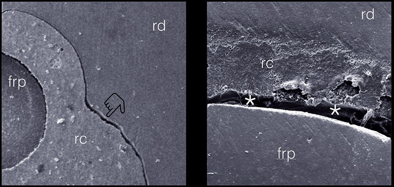
a. Pointer shows a breakdown between resin cement and root dentin. b. The asterisks show a debonding between a FRP surface and the resin cement (check on how to improve bonding between FRP and RC at our last text at BMC’s second edition - part I of this text). Different interfaces that needs different bonding approaches and its failures could result in clinical restorative failures. rd : root dentin ; rc : resin cement ; frp : fiber reinforced post.
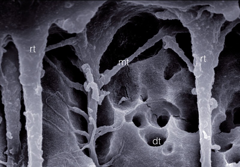
Basically, the challenge to bonding to root canal walls, are similar to bonding to coronal dentin, with a few more obstacles, like endodontic canal depth, tubular geometry, different dentin morphology (calcospherites – fig. 2) and non-regular surface demanding more time and attention to clinical subtle details.
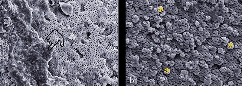
a. Images show a disrupted interface over calcospherite like interfaces, the pointer shows, adhesive/composite remnants over the dentinal surface. b. The calcospherites globules (asterisks) clearly seen at the pulpar floor dentin.
Nakabayashi et al [1], in 1982, published their classic paper on how resin infiltration of acid-etched dentin completely transforms the surface from one that is crystalline, acid-sensitive, and relatively hydrophilic to a structure that is organic, acid-resistant, and relatively hydrophobic. They showed that following acid etching and water rinsing, the mineral phase of dentin was removed. All that was left of the original dentin was the collagenous matrix. The solubilized…